
Sonography or Ultrasound imaging is a harmless and non-invasive medical test, which allows physicians to diagnose the medical condition of the patient without unnecessary pricking of the skin or piercing of the body parts.
What is Sonography (Ultrasound):
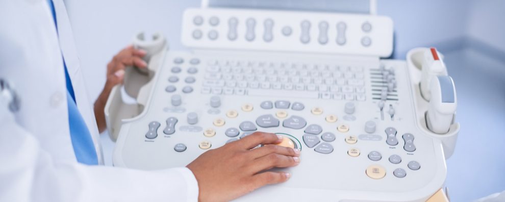
Ultrasound imaging, commonly called sonography involves exposing body parts to high-frequency sound waves to see images of the inner parts of the body. Examination by ultrasound, as opposed to X-rays, never involves the use of ionizing radiation. Images are captured in real-time.
Types Of Ultrasound:

The usual Sonography or Ultrasound examination is made with a transducer (ultrasound probe) on the skin. Although, sometimes the technicians and doctors can get a much better image by inserting the probe into one of the body’s natural openings:
- Transvaginal ultrasound: Inserting the probe into the vagina allows the examination of the uterus and the ovary
- Transrectal ultrasound: Inserting the probe into the rectum allows the examination of the prostate
- Transesophageal echo: Inserting the probe into the esophagus to allows the examination of the heart and the esophagus
Uses of Ultrasound imaging:
It helps in examine the internal body organs including:
- The liver
- The bladder
- The pancreas
- The eyes
- The spleen
- The heart
- Glands, like the thyroid and parathyroid
- The reproductive organs, including ovaries, uterus, and covering of the testicles (scrotum)
Ultrasound imaging can also be applied to guide needles into the body for obtaining biopsies.
Benefits and risks
Benefits
- Most ultrasound examinations are non-invasive (no needles or injections), meaning that they are painless.
- Ultrasound testing is less expensive, widely available and is very easy to carry out.
- Ultrasound Imaging never exposes you to the cell damage.
- Soft tissues that are hard to see on images from X-rays can be examined on ultrasound scans.
- With real-time imaging, an ultrasound is an excellent tool to use as a guide in minimally invasive procedures, such as needle aspiration and needle biopsies.
Risks
There are no reported harmful effects of a standard diagnostic ultrasound.
Limitations
- The bowel and any organ hidden by the intestine cannot be examined because gas or air blocks ultrasound waves.
- Scanning overweight patients can be challenging because of the fat tissue.
- Ultrasound waves cannot easily pass through the bones in the body.
Conclusion:
Ultrasound is a painless and non-dangerous diagnostic tool. It can help in examining internal body organs.
References:
- http://www.webmd.com/a-to-z-guides/what-is-an-ultrasound?page=2#1
- http://www.radiologyinfo.org/en/info.cfm?pg=genus
- http://www.nhs.uk/Conditions/Ultrasound-scan/Pages/Introduction.aspx